Research
The BioMC is supporting interdisciplinary and international collaborative projects integrating materials manufacturing, biomaterial synthesis/characterization, biomedical device design and clinical implementation.
Local Collaborations
Subcutaneous Vein Detection and Mapping for Home Hemodialysis Self-cannulation
Related Researchers: Prof. Kai Huang & Prof. Patrick Lee
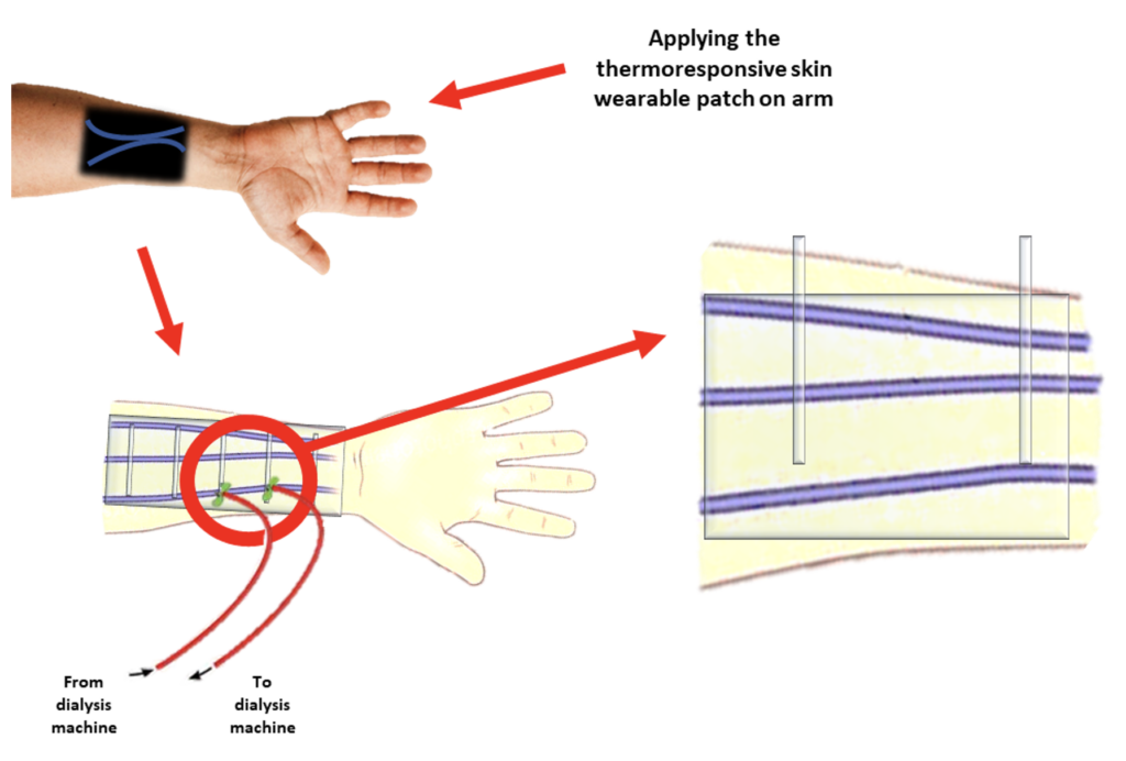
Although home hemodialysis (HHD) benefits renal patients including improving their lives’ quality, self-cannulation is currently a limiting and challenging step in their treatment. Some works have been performed to help patients locate their veins, such as surgical skin markers, near-infrared or ultrasound mapping; however, a more practical and easy-to-use solution without the need for external and expensive devices or permanent markings on the body has still remained unsolved. In this study, we aim to solve this problem by developing an epidermal film/patch fabricated from flexible and biocompatible polymer such as polydimethylsiloxane (PDMS) and thermochromic liquid crystals (TLCs) to visualize veins. The ability of TLCs to change color when adjacent to temperature difference of veins from the skin have made them suitable materials in the vein-visualizer epidermal patch. The patch simply contains a three-layered structure consisting of the thin PDMS substrate, embedded TLCs and the PDMS film as the encapsulant of the biomatrix composite on the top-layer. A customized formulation of TLCs is required in order to specifically be employed in this patch in the desired temperature operating of the forearm skin and vein temperature difference, 33-37 °C, and 0.5-1.2 °C, respectively. Spin coating and blade coating are the main processes for the fabrication of this polymer-based TLC-embedded matrix patch. Figure 1 is depicting a schematic of the final target of this project which is to reveal the forearm veins in home hemodialysis applications.

Schematic illustration of the thermoresponsive vein-mapping skin wearable patch.
Related Researchers: Prof. Kai Huang & Prof. Qian Lin
Nanomedicine has emerged as a promising tool for the treatment of various diseases, including cancer and neurodegenerative diseases. While some biomaterial-based nanomedicines have been approved for clinical applications, the potential risks of these medicines on brain function remain poorly understood. We are investigating the impact of nanomedicines on brain function using the zebrafish model. Zebrafish offer genetic tractability, large-scale behavior-based drug screening, and a small and transparent brain for brain-wide neural recordings. We use spontaneous and sensorimotor behaviors to assess brain function. We aim to provide solid data and insight into the impact of nanomedicines on brain function and inform the development of safe nanomedicines for the treatment of neurological diseases.
Methotrexate-loaded microbubbles for image-guided treatment of inflammatory bowel disease
Related Researchers: Prof. Kai Huang & Prof. Naomi Matsuura
Methotrexate (MTX) is a systemically injected immunomodulatory drug used to treat inflammatory bowel disease (IBD). In this project, we aim to develop ultrasound-responsive MTX-loaded, phospholipid microbubbles (MBs) with comparable in vitro stability as existing clinical MBs and with sufficient drug-loading for future ultrasound-guided, targeted, MTX treatment of IBD. MTX drug loading and stability, acoustic response and drug release of fluorocarbon gas-filled MTX-loaded MBs using different lipid shells will be assessed in comparison to plain MBs. Based on these results, MTX-loaded MBs will be evaluated for future, targeted delivery of MTX in preclinical models of IBD.

Visual overview of how MBs can be used as drug delivery systems upon injection into the vasculature of patients with IBD. As MBs reach vasculature of inflamed tissue, MBs stay intact and do not release loaded drug-cargo without US stimulation. Image courtesy of Yara Ensminger’s thesis.
International Collaborations
Comparing immersiveness and perceptibility of spherical and curved displays
Related Researchers: Prof. Mark Chignell & Prof. Hideki Koike (Tokyo Institute of Technology)
Curved displays are believed to create a feeling of immersiveness similar to virtual reality. However systematic studies are needed to demonstrate that this is, in fact, true and under what conditions. In an experimental study 24 participants compared five different displays (concave, convex, hemisphere, sphere and a flat display) in terms of their immersiveness and perceptibility, and they also rated their overall preferences. Both immersiveness and perceptibility affected overall preference ratings. Participants gave higher preference ratings to the convex and concave displays, which were rated high in immersiveness and perceptibility, but gave lower preference ratings to the hemisphere/spherical display which had high ratings on immersiveness but low ratings on perceptibility. The study results imply that curved displays do indeed create a feeling of immersiveness. Concave and convex displays were rated highly favorably and should receive more attention for applications where visual experience and a feeling of immersiveness is particularly important.
Related publication: Urakami, J., Matulis, H., Miyafuji, S., Li, Z., Koike, H., & Chignell, M. (2021). Comparing immersiveness and perceptibility of spherical and curved displays. Applied Ergonomics, 90, 103271.

The Five displays used in the Experiment.
Related Researchers: Prof. Patrick Lee & Prof. Hiroaki Takehara (University of Tokyo)
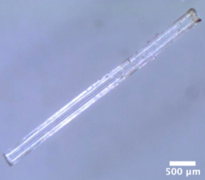
The sustainably sourced and biocompatible polyester, poly(L-lactide) (PLLA), has found increasing use in applications ranging from food packaging to textile fibers. Additionally, its biocompatibility makes it ideal for use in biomedical applications including micro needles for both transdermal drug delivery and sensing applications. Unfortunately, its modulus and strength are insufficient for such applications. This challenge can be overcome by blending PLLA with poly(D-lactide) (PDLA), a combination forming stereocomplex crystallites (SCs) with a melting temperature 50 °C above that of PLLA homocrystals. These SCs are intrinsically stiff and strong, and promote matrix crystallization, both translating to improved composite mechanical properties. However, SC morphology and topology determine composite properties. Herein, we explore the effects of blend composition and processing conditions on SC morphology and topology within a PLLA matrix. Moreover, we explore structure – property relationships in the fabrication of high-aspect ratio microneedles prepared via a novel vacuum-assisted micro molding approach. By incorporating SCs in this micro molding approach, we alter microneedle flexural properties, targeting reduced deformation during injection.

Fabricated microneedle.
Related Researchers: Prof. Naomi Matsuura, Prof. Alexander Eggeman (University of Manchester) & Prof. Sarah Cartmell (University of Manchester)
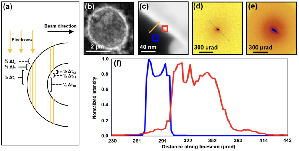
Microbubbles are clinical ultrasound contrast agents comprised of a high density, inert gas core generally stabilized by a surrounding shell of biocompatible phospholipids. Beyond diagnostic purposes, ultrasound-stimulated microbubbles have garnered significant interest for drug delivery applications owing to the therapeutically relevant bioeffects they can elicit depending on the ultrasound parameters. By directly loading the drug of interest on the lipid shell, not only may the narrow therapeutic index of its respective commercial formulation be improved, but it can offer a localized release of the drug directly to the targeted lesion in a non-invasive, spatiotemporally controlled manner. While a plethora of drug-loaded bubbles have been investigated in the literature, the direct confirmation of the presence of drug crystals (or molecules) within the stabilizing shell has yet to be examined. To investigate this, 4D-scanning transmission electron microscopy (4D-STEM) may be employed to directly examine the local physical features of such drug-loaded bubbles and dynamically acquire spatially-resolved electron diffraction patterns of the drug. Successful identification of the drug in the phospholipid shell will not only provide direct evidence of drug-loaded bubbles on the molecular scale but assess the homogeneity of drug distribution within the shell as well as the possibility of bubble size-dependent drug loading.

(a) Schematic of a geometric model representing sample thickness changes (Δtn) along the electron scanning beam in x-direction (n represents integers); (b) high annular angle darkfield (HAADF) scanning transmission electron microscopy (STEM) image of a silica-coated phospholipid microbubble; (c) illustration of two scanning points on the exterior of a bubble: (24, 48) in blue and (35, 37) in red with the direction of the scanning direction shown in yellow; diffraction patterns of scanning points at (d) (24, 48) and (e) (35, 37); and (f) electron distribution histogram across diffraction pattern linescan of the two scanning points. Phospholipid-stabilized microbubbles were coated in a silica gel by the Stöber method by using silica as a control material.
Related Researchers: Prof. Naomi Matsuura, Prof. Marco Domingos (University of Manchester)
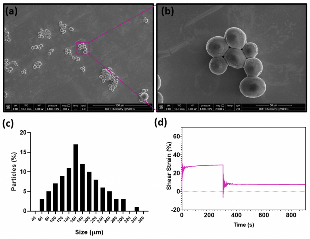
Osteoarthritis (OA) affects millions globally, with knee OA accounting for the majority of these cases. Patients affected by the disease undergo significant pain symptoms and experience a decline in their quality of life (QoL), while the condition itself is marked by cartilage degradation, alterations in subchondral bone, and synovial inflammation. Genicular artery embolization (GAE) has emerged as a promising treatment option, involving the administration of an embolic agent into the genicular arteries to impede neovessel blood flow and alleviate angiogenesis-associated symptoms. However, current embolic agents lack GAE-specific indications, highlighting the need for the development of a tailored embolic agent for OA treatment. To bridge this gap, sodium citrate (Cit)-based hyaluronic acid (HA) particles (HACit) have been synthesized, with HACit-10 demonstrating favorable properties, including a microspherical shape, biodegradability, and adequate sizing distribution. Further in vitro characterization of these particles will be performed using nuclear magnetic resonance (NMR) to assess the bonding regime between Cit and HA, as well as creep recovery tests to evaluate the viscoelastic behavior and long-term mechanical properties of the particle formulation. These analyses, conducted in collaboration with the University of Manchester, will inform the subsequent utilization of these particles in pre-clinical animal models, furthering our understanding of their potential in OA treatment.

(a) and (b) Representative scanning electron microscopy (SEM) of HACit-10. (c) histogram representing the size distribution of HACit-10. (d) Creep recovery curve demonstrating the time-dependent deformation and recovery behavior of HACit.
